
Sebaceous cyst — diagnosis, drainage and surgical removal at SkinWise Clinic, Bengaluru.

By · Dermatologist, SkinWise Clinic Published Last reviewed

Sebaceous cyst: when a small lump calls for gentle attention

A patient feels a soft, round lump under the skin on the back, the scalp, behind the ear. It doesn’t hurt. It moves slightly when pressed. It’s been there for months — sometimes years — and either it’s growing imperceptibly or it just suddenly announced itself by becoming sore.

This is almost always a sebaceous cyst (or, more accurately, an epidermal cyst — but more on that in a moment). They’re among the most common skin growths we see at SkinWise. They’re benign. And they’re straightforward to treat if you don’t make any of the easy mistakes — squeezing, picking, half-draining, or leaving an inflamed one until it bursts.

Here’s how we think about them, when to leave them alone, and when removal is the right call.

What it actually is

“Sebaceous cyst” is the common phrase, but most of these lumps are technically epidermal cysts (or epidermoid cysts, or epidermal inclusion cysts — three names for the same thing). They form when skin cells get trapped in a small sac under the surface and keep producing keratin — the same protein that makes up the outer layer of skin and the inside of hair shafts.

The result is a slow-growing, dome-shaped lump filled with thickened, cheesy, sometimes faintly malodorous material. It’s sealed inside a thin sac (the cyst wall). Most have a tiny visible central pore — the punctum — through which they occasionally leak.

A few important relatives, often confused with epidermal cysts:

  • Pilar cysts — almost always on the scalp; very similar treatment but the wall is structurally different
  • Sebaceous cysts (true) — actually arise from sebaceous glands, contain oily sebum, much rarer than common usage suggests
  • Lipomas — soft, mobile fatty tumours; feel different (rubbery, deeper, no overlying pore)
  • Abscesses — acute, red, painful, hot; different problem with different urgency
  • Dermoid cysts — congenital, often on the face/scalp in a midline position; usually present from childhood

Telling them apart matters because some change the surgical approach. A dermatologist examining the lump can distinguish them in most cases at the bedside; in unusual locations or atypical presentations we may image (ultrasound) before excising.

What it looks and feels like

  • A round, dome-shaped lump under intact skin
  • Soft to firm; movable; doesn’t feel “stuck” to deeper structures
  • A few millimetres to several centimetres
  • Skin-coloured or slightly yellowish-white
  • Often a small central black dot or pore (the punctum)
  • Usually painless
  • Occasionally tender if recently irritated or about to flare
  • May leak a thick, pasty, foul-smelling substance if disturbed

Common sites: scalp, behind the ears, neck, upper back, chest, the area below the jaw, and the genital region. They can occur anywhere there’s hair-bearing skin.

What causes them

  • Blocked hair follicle / damaged skin cell line. The most common origin.
  • Skin trauma or surgical wound. Skin cells trapped under the surface during healing can become a small cyst over months.
  • Acne-prone or oily skin. Higher background prevalence.
  • Genetic predisposition. Some patients have multiple cysts over a lifetime; family history is common.
  • Conditions associated with multiple cysts. Gardner’s syndrome (rare familial condition) is associated with multiple epidermal cysts plus colon polyps — flagged when the history suggests it.

They are not caused by:

  • Poor hygiene
  • Diet
  • “Bad” bacteria
  • “Toxins”

Are they dangerous?

In the overwhelming majority of cases — no. Sebaceous/epidermal cysts are benign and have essentially no malignant potential. The vanishingly rare exceptions (proliferating trichilemmal tumour on the scalp, malignant transformation in long-standing cysts) are unusual enough that they shouldn’t change the overall calculus.

What does warrant attention:

  • Sudden growth
  • Persistent pain, redness, warmth (suggests infection or impending rupture)
  • A cyst that has ruptured under the skin
  • Repeated infections in the same site
  • Atypical features that don’t fit the usual cyst (firm, fixed, ulcerated)
  • A cyst in a sensitive or functionally important area (eyelid, genital, near a joint)

When to leave it alone

A small, painless, slow-growing cyst in an asymptomatic area can absolutely be left alone. Removal is elective, not urgent.

When we generally recommend leaving them:

  • Very small, stable, in a non-cosmetically-bothersome location
  • Patient is not bothered, and we’ve confirmed the diagnosis
  • Pregnancy or breastfeeding (defer to after, unless infected)

When removal is the right call

We recommend removal when:

  • The cyst is growing, large, or visibly distressing
  • It’s been infected once — repeat infections are common, and each one makes the surgery slightly more complex
  • It’s in a high-friction area where it gets repeatedly irritated (waistband, bra-line, collar)
  • It’s leaking or has burst recently
  • The patient simply wants it gone and the location and timing are reasonable
  • Diagnosis is uncertain and tissue is needed to confirm

Our approach at SkinWise

Step 1: confirm the diagnosis

A focused examination, a look for the classic punctum, sometimes ultrasound for cysts in unusual locations or for accurate sizing in deeper sites.

Step 2: handle infection first

An inflamed, infected cyst is not the right setting for definitive removal. Cutting into an infected cyst increases the risk of incomplete removal and worse scarring.

For an inflamed cyst:

  • A short course of oral antibiotics
  • Warm compresses
  • Sometimes intralesional steroid injection to reduce inflammation
  • Incision and drainage if there’s frank abscess formation — this is temporary; the cyst wall remains and we plan definitive removal 6–8 weeks later

Step 3: definitive surgical removal

Once the cyst is calm, definitive removal is a short outpatient procedure under local anaesthetic:

  1. Local anaesthetic injected around the cyst
  2. Small incision over the cyst, planned along skin-tension lines for best scar
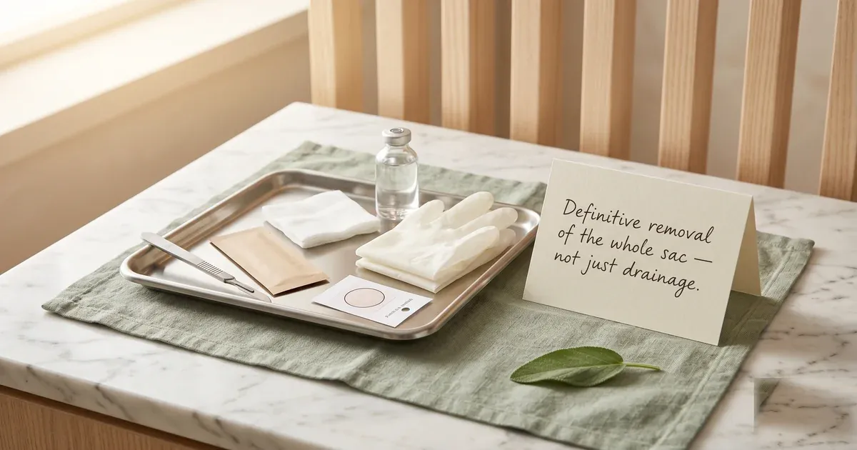
  3. Complete removal of the cyst wall — this is the part that prevents recurrence. Leaving any sac behind is the main reason cysts come back.
  4. Sutured closure in layers
  5. Dressing and aftercare brief

The whole procedure takes 20–45 minutes depending on size and location. Patients walk out the same day.

Step 4: aftercare

  • Mild soreness for 1–2 days; paracetamol as needed
  • Keep the area clean and dry; dressing change as instructed
  • No swimming or heavy sweating until sutures are out
  • Suture removal at 5–10 days depending on location (scalp and back later than face)
  • Silicone scar gel or sheet often recommended from 2 weeks
  • Final scar settling over 3–6 months — usually a thin line, easily concealed

What not to do

  • Don’t squeeze it. Pressure forces the contents into surrounding tissue and triggers inflammation — sometimes infection. Cysts squeezed in front of bathroom mirrors are how a quiet 1-cm lump becomes a 4-cm red painful one in 48 hours.
  • Don’t try to pick out the “core.” You can extract some keratin through the punctum and it briefly looks smaller — the sac is still there, and the cyst always comes back larger.
  • Don’t go to a salon to have it “removed.” Salon facial extractions of cysts are unsterile, incomplete, and a leading cause of infection.
  • Don’t take repeated short antibiotic courses without addressing the cyst. Each course suppresses an inflamed cyst briefly but doesn’t change the underlying anatomy. Definitive removal is what closes the loop.
  • Don’t apply Ayurvedic pastes or herbal poultices to an inflamed cyst. Heat, irritation and skin pigmentation often follow, with no benefit.
  • Don’t panic about cancer in a typical-looking benign cyst. Examination usually reassures; biopsy of removed tissue confirms when there’s genuine ambiguity.

Frequently asked questions

Will it come back? If the complete sac is removed — usually no. If it was incised and drained but the sac left behind — yes, often within months. We always aim for complete excision.

Will there be a scar? A thin line scar in the direction of the natural skin lines. Usually fades to near-invisible at 6–12 months with good aftercare. Larger cysts and high-tension areas (chest, shoulders) leave more noticeable marks; we discuss expectations upfront.

Can it become cancer? Essentially no. The rare exceptions are unusual scenarios and are picked up on examination or histology of the removed cyst.

Why does it smell? The keratin and sebum inside have been broken down by skin flora. The smell is a feature of the cyst contents, not an infection — though infection makes it worse.

Why is it suddenly sore? Most commonly because the cyst has briefly leaked into surrounding tissue, triggering an inflammatory response. This is sometimes mistaken for an acute infection — sometimes it is one, sometimes it’s purely inflammatory.

Should I just drain it at home? No. Home drainage almost always leaves the sac behind, often gets infected, and leaves a larger and more difficult cyst to remove properly.

Can it spread elsewhere? A single cyst doesn’t “spread” — but patients prone to cysts may develop new ones independently. We sometimes counsel skin care and trigger reduction in those patients.

My child has one — should we remove it? Cysts in children are rarer; congenital cysts (dermoid cysts) sometimes get confused with epidermal cysts. We examine, sometimes image, and then decide. Many can wait until later childhood; some need earlier removal for functional or diagnostic reasons.

Where to go from here

If you’ve had a lump under the skin for more than a few weeks, or you’ve had one drained that keeps coming back, book a consultation. We’ll examine, confirm the diagnosis, decide whether removal is the right call, and schedule it at a time that suits you.

Related reading:

Book a consultation with Dr Khushboo

Book consultation